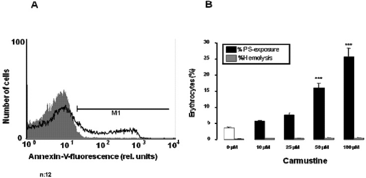
Figure 3.
Effect of carmustine on PS exposure and hemolysis. (A) Original histogram of annexin V binding of erythrocytes following exposure for 48 h to Ringer solution without (grey shadow) and with (black line) presence of 100 µM carmustine; (B) Arithmetic means ± SEM (n = 12) of erythrocyte annexin V binding following incubation for 48 h to Ringer solution without (white bar) or with (black bars) presence of carmustine (10–100 µM). For comparison, arithmetic means ± SEM (n = 4) of the percentage of hemolysis is shown as grey bars. *** (p < 0.001) indicates significant differences from the absence of carmustine (ANOVA).

