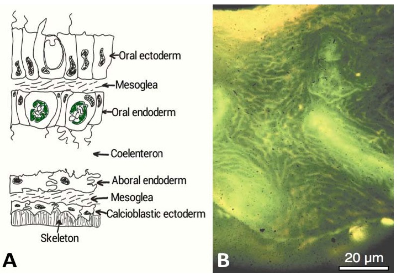
Figure 2.
(A) A cross-sectional diagram through the living tissue portion of a coral and showing the interface with the exoskeleton. The calcioblastic epithelium secretes the organic matrix probably embedded within intracellular vesicles into micron or nanometric sized spaces. Biological control of mineralization is strongly implicated in corals in other ways. There are semi-permeable tight septate junctions between the cells that control ion transport and other molecules according to size and charge (After Allemand et al. [22]). The calcioblastic epithelium effectively facilitates the laying down of the inorganic skeleton; (B) Acridine orange staining of the organic matrix of Acropora sp. skeleton, which appears strong yellow at the growing region and green deeper inside the skeleton. The pale yellow regions are the centres of calcification. This microscope image was taken under polarized light and shows the global distribution of intra-skeletal organic matrices throughout the entire skeleton [25]. The coral tissue has been removed to view this organic matrix. (Reproduced with permission from the Institute of Paleobiology, Polish Academy of Sciences, Gautret et al. 2000 [25]).

