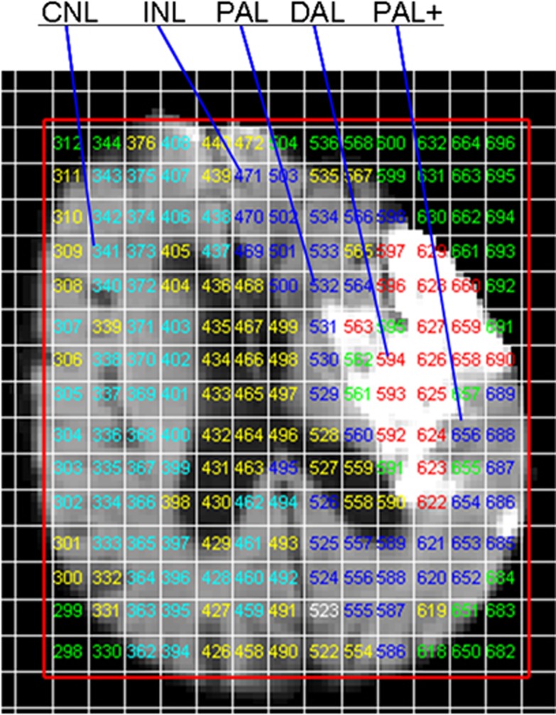
Figure 1.
Example of voxel grid categorization according to tissue appearance on diffusion tensor imaging (DTI). CNL, contralateral normal brain; DAL, definitely abnormal tissue; INL, ipsilateral normal brain; PAL, possible abnormal tissue; PAL+, tissue one voxel thick immediately outside the lesion.

