Figure 1.
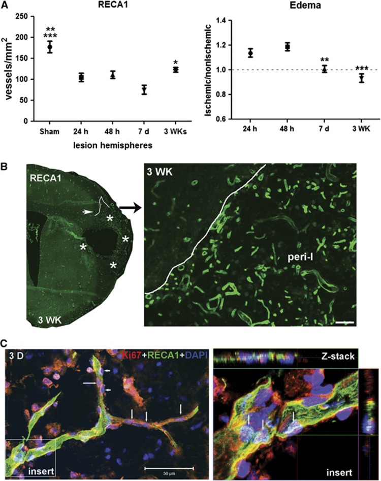
Increased number of newly formed vessels in ischemic hemispheres at 3 weeks of reperfusion. (A) Graph showing number of RECA1 staining microvessels in sham-operated and ischemic hemispheres. Focal ischemic stroke with reperfusion injury resulted in loss of RECA1-positive microvessels in ischemic hemispheres at 24 and 48 hours and 7 days. At 3 weeks after stroke, the number of RECA1-positive microvessels was increased compared with that at 7 days. *P<0.05 vs. 7 days, **P<0.01 vs. 48 hours and 3WK; ***P<0.001 vs. 24 hours and 7 days, n=6 per group. The edema in ischemic hemispheres at 24 hours, 48 hours, 7 days, and 3 weeks of reperfusion compared with nonischemic hemispheres expressed as edema index. **P<0.01, ***P<0.001 vs. 24 and 48 hours, respectively, n=6 per group. The number of vessels was normalized for edema. (B) Immunohistochemistry of RECA1, a marker of endothelial cells, represents microvessels in peri-infarct area in ischemic hemisphere with reperfusion of 3 weeks (3WK). Lines outline the border of lesion areas. Stars indicate the peri-infarct area. Right panel shows the peri-infarct (peri-I) area in higher magnification. A higher density of microvessels was seen in the peri-infarct area. Scale bar=50 μm. (C) Three-dimensional (3D) confocal microscopy shows that RECA1-positive vessel (green) contains nuclei expressing Ki67 (red, arrows), a marker of cell proliferation, in peri-infarct area at 3 weeks after stroke. Arrowheads show pericyte-like cells expressing Ki67. 4′-6-diamidino-2-phenylinidole (DAPI) (blue) was used to stain nuclei. Z-stack confocal micrograph (insert) shows colocalization of Ki67 with DAPI in vascular endothelial cells (arrows). Scale bar=50 μm.

