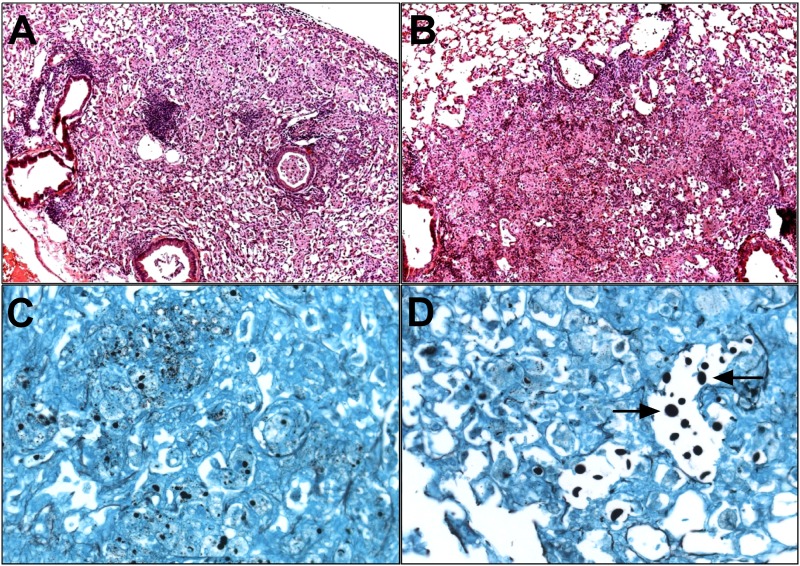
FIG 7 .
Lungs of XID mice exhibit disorganized inflammation and increased numbers of large, extracellular C. neoformans yeast in lung sections. (A to D) Representative images of H&E-stained lung section (magnification, ×10) from CBA/CaJ (A) and XID mice (B) 6 weeks after infection with C. neoformans strain 52D and Gomori methenamine silver-stained section (magnification, ×40) from CBA/CaJ controls (C) and XID lungs (D) at week 6 (n = 3). The arrows in panel D point to enlarged, extracellular yeast.

