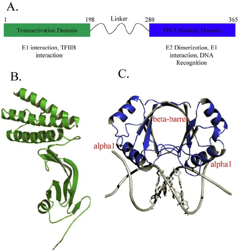
Figure 1.

A) Schematic of the HPV16 E2 gene and known proteins of interaction. B) Ribbon diagram of the structure of the HPV16 E2 transactivation domain (31) .C) A ribbon representation of the structure of the HPV18 E2 DNA binding domain showing the dimeric protein bound to the DNA sequence ACCGAATTCGGT (PDB-ID: 1JJ4). The recognition helix alpha1 makes direct contact with the major groove of the DNA. The bases of the spacer region AATT are depicted.
