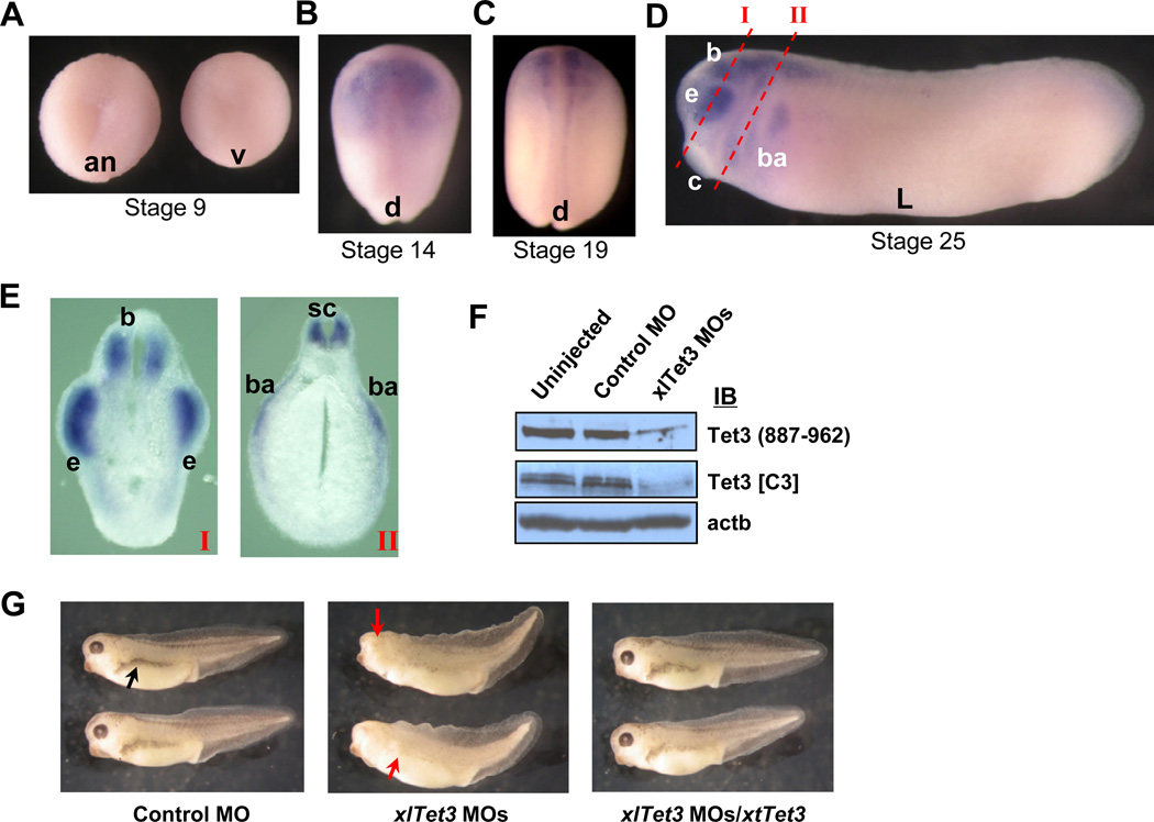
Figure 1. Tet3 is important for early eye and neural development.
(A–E) Spatial expression profile of xlTet3 by in situ hybridization at stage 9 (A), 14 (B), 19 (C) and 25 (D and E). The sites of sections I and II in panel E are noted by red dashed lines in panel D. Animal view (an), vegetal view (v), dorsal view (d), lateral view (L), brain (b), eye (e), cement gland (c), branchial arches (ba) and spinal cord (sc).
(F) Western blot showing depletion of endogenous Tet3 protein by xlTet3 MOs in stage 14 embryos.
(G) Developmental defects in stage 35 embryos caused by Tet3 depletion. Small head, eyeless and missing pigmentation phenotypes in xlTet3 MOs-injected embryos are noted by red arrows, and the normal pigmentation in control embryos is noted by a black arrow.
See also Figure S1.

