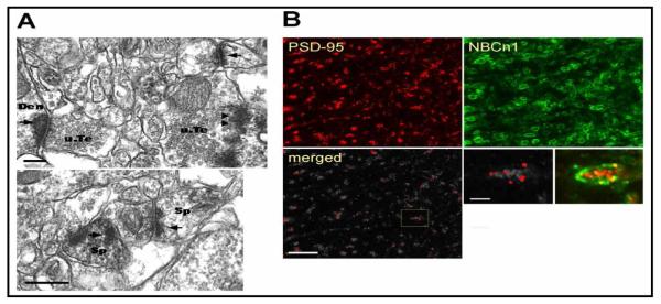
Fig. 1.

Localization of NBCn1 at synapses in hippocampal CA3 neurons. A) Electron micrographs of NBCn1 immunope-roxidase in dendrites. NBCn1-positive dendrites contacted by unlabeled nerve terminals are shown. Dendritic spines positive to NBCn1 are also indicated. NBCn1 stainings are mostly in postsynaptic membranes (arrows) but often found in presynaptic stainings (arrow heads). Den, dendrite; Sp, spine; u.Te, unmyelinated terminal. Bar: 0.25 μm. B) Double-label immunofluorescence of NBCn1 and PSD-95 in CA3 pyramidal neurons. Alexa 488-labeled NBCn1 (green) and Alexa 594-labeled PSD-95 (red) were used. Images were taken in 100 × magnification (bar: 10 μm). The areas, where the two immunofluorescence signals were merged (white areas), were identified using ImageJ software with the colocalization finder plug-in.
