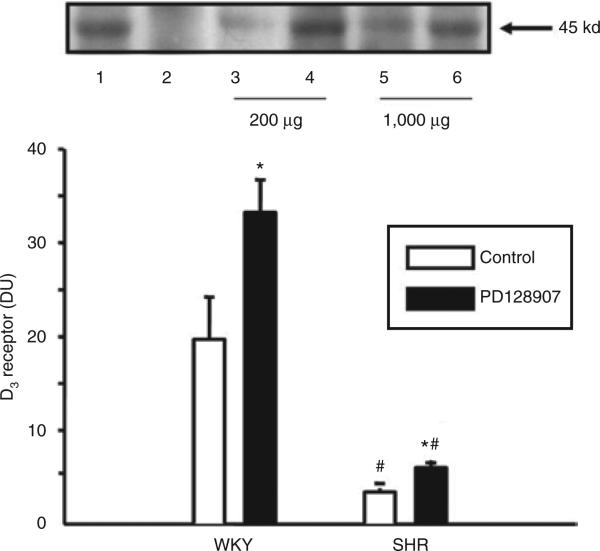
Figure 6.
Effect of a D3 receptor agonist, PD128907, on the co-immunoprecipitation of D3 and ETB receptors in WKY and SHR RPT cells. The cells were incubated with PD128907 (10–7 mol/l) for 24 h. Thereafter, the samples were immunoprecipitated with an anti-ETB receptor antibody and immunoblotted with an anti-D3 antibody (*P < 0.05 vs. control, #P < 0.05 vs. WKY n = 8/group, analysis of variance, Duncan's test). One immunoblot (45 kd) is depicted in the upper panel: (lane 1, positive control; lane 2, negative control; lane 3, vehicle-treated RPT cells from WKY rats; lane 4, PD128907-treated RPT cells from WKY rats; lane 5, vehicle-treated RPT cells from SHRs; lane 6, PD128907-treated RPT cells from SHRs). For a positive control, anti-D3 antibodies (1 μg/ml) were used as the immunoprecipitant; for a negative control, normal rabbit IgG (1 μg/ml) was used as the immunoprecipitant, instead of the anti-ETB antibodies, and immunoblotted with anti-D3 antibodies as above. The amount of D3 receptor antibody needed to immunoprecipitate ETB receptors in SHR RPT cells (1,000 μg, lanes 5 and 6) is five times that used in WKY RPT cells (200 μg, lanes 3 and 4). RPT, renal proximal tubule; SHR, spontaneously hypertensive rat; WKY, Wistar-Kyoto.

