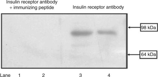
Figure 1.
Insulin receptor expression in HEK293 cells. Cell lysate proteins (50 μg) from RPT cells from WKY rats (lanes 1 and 3) and HEK293 cells (lanes 2 and 4) were subjected to immunoblotting with anti-insulin receptor antibody (1:250). In HEK293 cells and RPT cells from WKY rats, the 95-kDa band was no longer visible when the antibody was preadsorbed with the immunizing peptide (1:10 wt/wt incubation for 12 h). The molecular sizes are indicated. RPT, renal proximal tubule; WKY, Wistar-Kyoto.

