
Figure 3.
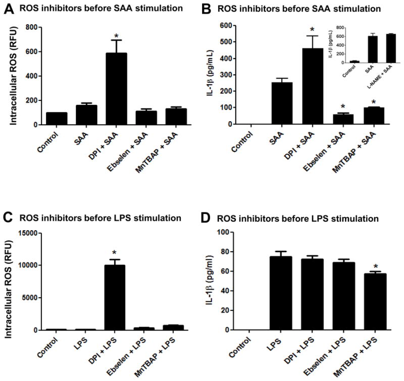
Effect of ROS inhibitors added prior to SAA or LPS stimulation on IL-1β secretion and intracellular ROS levels. Primary mouse peritoneal macrophages (A, B) or transformed mouse peritoneal macrophages (C, D) were left to adhere for at least 24h. Flavoprotein inhibitor DPI (10 μM), ebselen (10 μM) or the MnSOD mimetic MnTBAP (100 μM) were added for 30 min prior to SAA (1 μg/mL) or LPS (100 ng/mL) stimulation, respectively. After 24h of simultaneous treatment, intracellular ROS levels were analyzed as described in Fig. 2 (A, C). IL-1β secretion was quantified by ELISA (B, D). The data are representative of at least two independent experiments. * = p < 0.05 compared to SAA (A,B) or LPS (C, D).