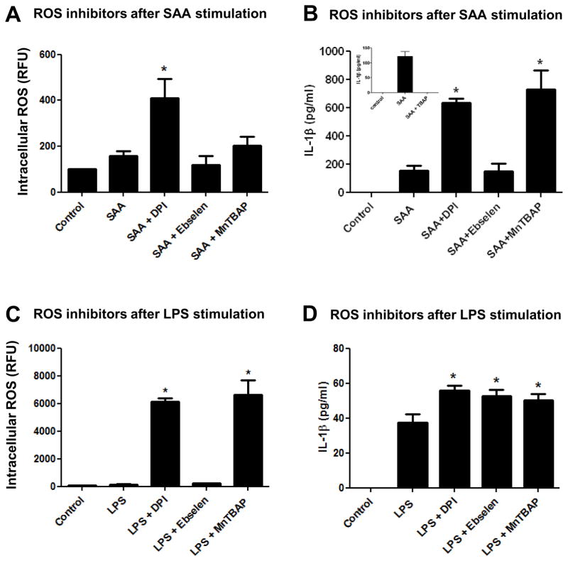
Figure 4.
Effect of ROS inhibitors added after SAA or LPS stimulation on IL-1β secretion and intracellular ROS levels. Primary mouse peritoneal macrophages (A, B) or transformed mouse peritoneal macrophages (C, D) were left to adhere for at least 24h and stimulated with SAA (1 μg/mL) or LPS (100 ng/mL) respectively for 8h. DPI (10 μM), ebselen (10 μM), MnTBAP (100 μM) or respective control (TBAP 100 μM, insert) were added to stimulated cells for another 16h. After a total 24h of simultaneous treatment, intracellular ROS levels were analyzed as described in Fig. 2 (A, C) and IL-1β secretion was quantified by ELISA (B, D). The data are representative of at least two independent experiments. * = p < 0.05 compared to SAA (A, B) or LPS (C, D).

