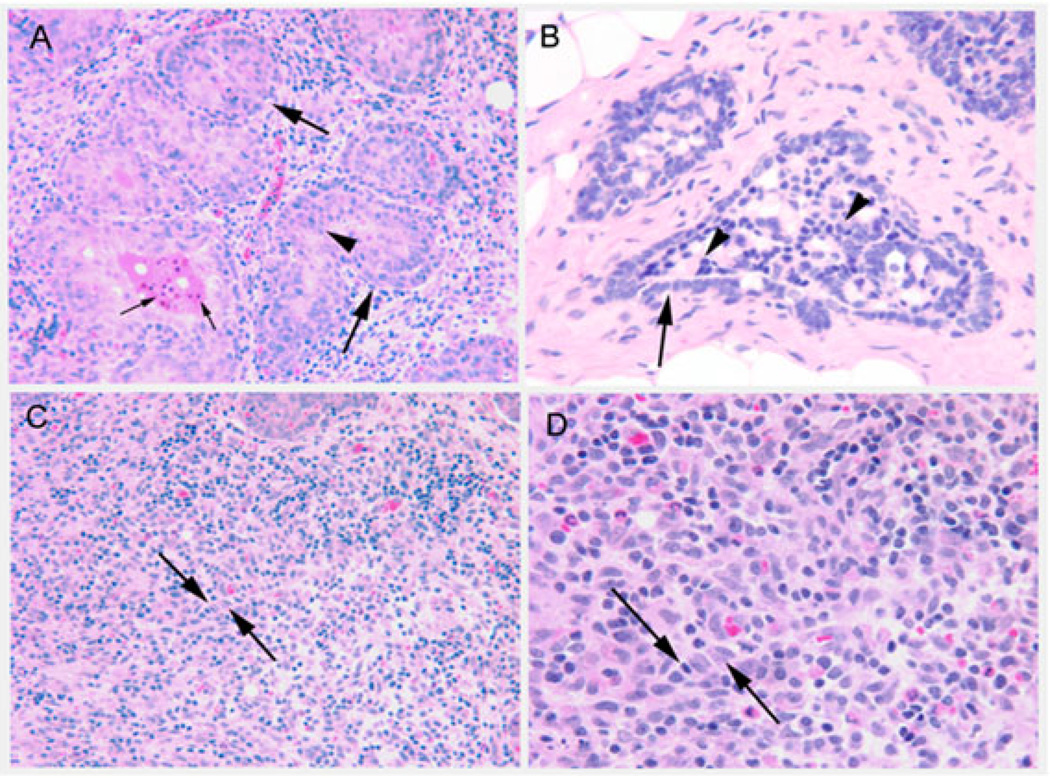
Figure 3.
Images of tumor phenotypes. Panel (A): Lobular carcinoma in situ, showing multilayered, nonpolarized epithelial cells (arrowhead) inside lobular ductules (large arrows). Small arrows indicate apoptotic bodies in the lumen of a lesion. Original magnification with 20× objective. Panel (B): Ductal carcinoma in situ, showing multilayered nonpolarized epithelial cells in a cribriform arrangement (arrowheads) within the lumen of a ductule (arrow). Original magnification, 40× objective. Panel (C): Invasive lobular carcinoma, with epithelial cells arranged in a single file embedded within the interstitial stroma (between arrows). Original magnification, 20× objective. Panel (D) enlargement of panel (C) showing nuclear heterogeneity within the invasive lobular carcinoma lesion (between arrows). Original magnification, 40× objective

