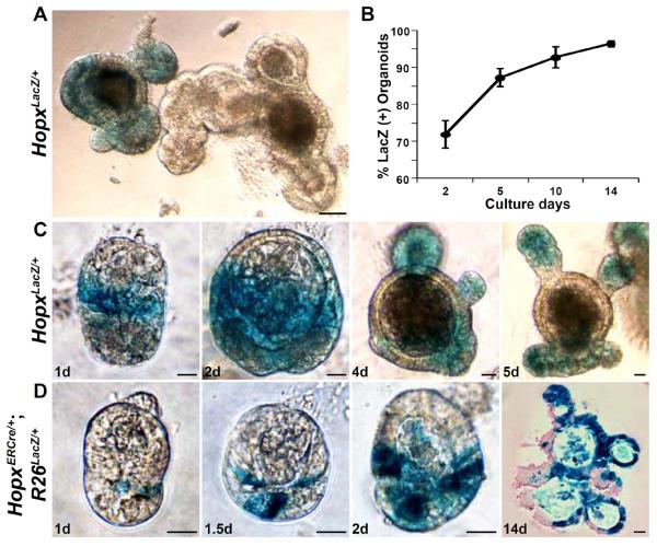
Fig. 2. Organoid cultures of Hopx-labeled cells.
(A) X-gal stained organoids from HopxLacZ/+ mice after 9 days of crypt culture. (B) Time course of the percentage of LacZ-positive organoids derived from HopxLacZ/+ mice. Two hundred organoids from three different HopxLacZ/+ mice were analyzed (error bars: 1 s.d.). (C) Examples of crypt organoids from HopxLacZ/+ mice stained with X-gal. (D) Examples of crypt organoids from HopxERCre/+;R26LacZ/+ mice (tamoxifen pulse for 12h on day 0.5 of culture). X-gal stained images are shown with a 14 day eosin counter-stained image. Scale bars: 50 μm (A) and 20 μm (C and D).

