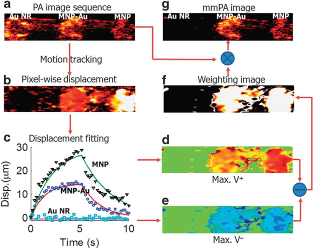
Figure 5.
Data processing in mmPA imaging of MNP-gold hybrid NPs [19]. (a) A conventional PA image of the phantom in Figure 4. (b) Maximum displacement resulting from the action of the magnetic field presented on a pixel – pixel basis. (c) Three representative displacement traces and their fitted curves over the entire time interval of the experiment for pixels in different inclusions. (d, e) Velocity computed over the full interval when the magnetic field is turned on and off. (f) Weighting image based on the magnitude of the difference between peak positive velocity in the first half and peak negative velocity in the second half. (g) mmPA image produced from the product of (a) with (f).

