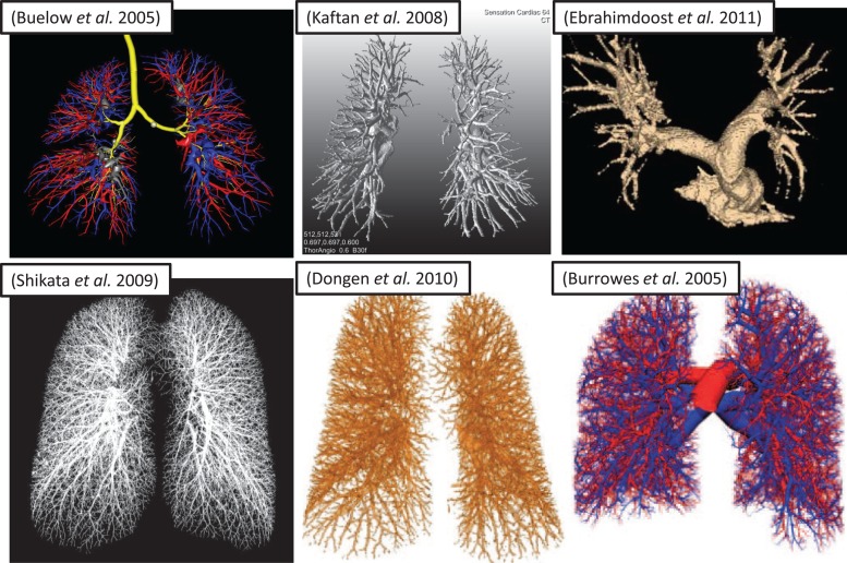
Fig. 3.
Resulting arterial trees, reconstructed using different automated and manual techniques from in vivo human volumetric CT scans. Different degrees of fine structure reconstruction are due, in part, to differences in image resolution: [65]-Buelow (voxel dimensions not given); [66] Shikata (0.6 × 0.6 × 1.3 mm); [67] Kaftan (0.6 × 0.6 × 0.6 mm); [68] Dongen (submillimeter, isotropic, but not specified); [69] Ebrahimdoost (0.66 × 0.66 × 1.0 mm); [70] Burrowes (0.68 × 0.68 × 1.4 mm).

