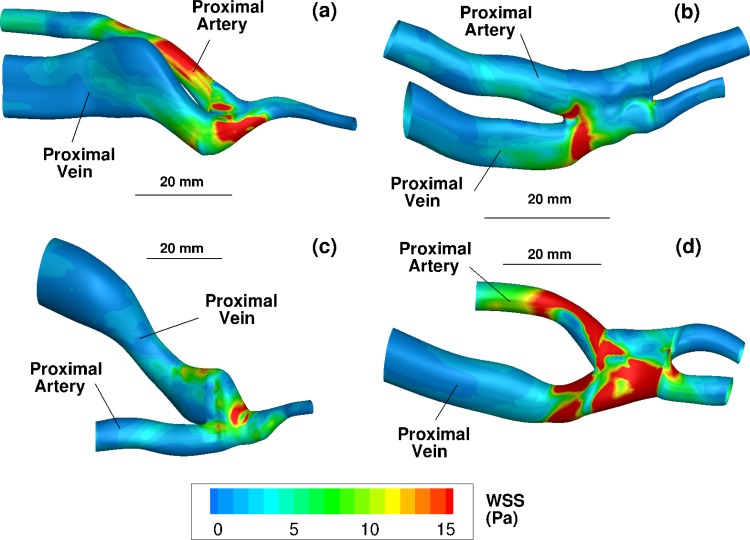
Fig. 1.
Three-dimensional ultrasound reconstructions of the four fistulae with the lumen colored by time-averaged wall shear stress (Eq. (3)) in Pa. (a), (b), (c), and (d) Reconstructions from patients 1, 2, 3, and 4, respectively. The view in each subfigure is shown from the skin toward the fistula. The bar labeled 20 mm shows the relative size of each figure.

