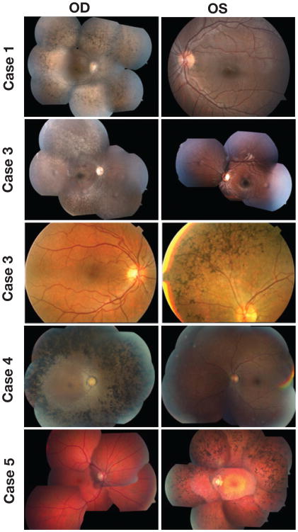
Fig. 1.
Color fundus photographs of both eyes of each patient showing typical retinitis pigmentosa (RP) abnormalities, including attenuated retinal vessels, intraretinal clumps of black pigment, and loss of retinal pigment epithelium in the affected eye of each patient; the fellow eye of each case presents a normal aspect. Color fundus images show typical RP changes and swelling at the optic disc area of the right eye of patient 1; affected right eye of patients 2 and 4 and typical RP abnormalities in the left eye of patients 3 and 5.

