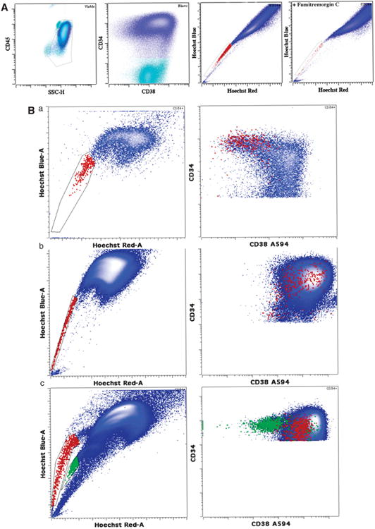
Figure 1.

Flow cytometry analysis of SP phenotype in relation to CD38 expression. (A) Typical example of FumC-mediated inhibition of SP phenotype in AML. CD34+ blasts first gated based on CD45 and side scatter and then on CD34 expression (shown in blue) demonstrated a SP of 0.55% (shown in red). When FumC was added to the sample, the proportion was reduced to 0.03% with identical gating. Dead cells were excluded using 7-amino-actinomycin D uptake prior to gating. (B) (a) Some AML cases demonstrate mild decrease of CD38 expression on SP blasts. SP blasts are depicted in red and bulk blasts are shown in blue. (b) The majority of AML cases exhibit bright CD38 expression. SP blasts are depicted in red and bulk blasts are shown in blue. (c) A single case demonstrated two distinct blast populations of different ploidy, one derived from the leukemic clone and one from normal population. The population with brighter Hoechst blue and red staining (higher ploidy) demonstrated SP (red) primarily with brighter CD38. Both the SP and the higher ploidy population demonstrated numerous AML-related genomic abnormalities. In contrast, the population with lower ploidy showed a SP (green) that had dim CD38 and did not contain any detectable cytogenetic abnormalities suggestive of residual normal blasts.
