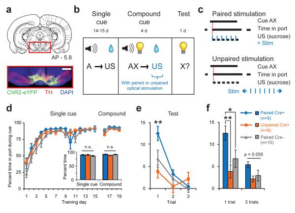
Fig. 2. Dopamine neuron stimulation drives new learning.
(a) Example histology from a Th::Cre+ rat injected with a Cre-dependent ChR2-containing virus. Vertical track indicates optical fiber placement above VTA. Scale bar = 1mm. (b) Experimental design for blocking task with optogenetics. All groups received identical behavioral training according to the “blocking” group design in Fig. 1a. (c) Optical stimulation (1s train, 5ms pulse, 20 Hz, 473nm) was synchronized with sucrose delivery in Paired (Cre+ and Cre–), but not Unpaired (Cre+), groups. (d) Performance across all single cue and compound training sessions. Inset, no group differences over last four days of single cue training or during compound training. (e) Performance during visual cue test. The PairedCre+ group exhibited increased responding to the cue relative to both control groups at test on the first trial (**p<0.005). (f) Visual cue test performance for the first trial and all three trials averaged. The PairedCre+ group exhibited increased cue responding relative to controls for the 1-trial measure (PairedCre+ vs. UnpairedCre+, **p=0.005, PairedCre+ vs. PairedCre–, *p=0.025, PairedCre– vs. UnpairedCre+,p=0.26); there was a trend for a group effect for the 3-trial average (main effect of group, p=0.055).

