Figure 2.

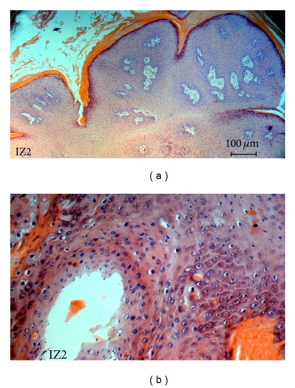
(a) Histopathology of a wart biopsy: detailed aspect of the IZ2 lesion exhibiting characteristic hyperkeratosis, acanthosis and dermal proliferation, indicated by arrows (100x). (b) Presence of koilocytosis.

(a) Histopathology of a wart biopsy: detailed aspect of the IZ2 lesion exhibiting characteristic hyperkeratosis, acanthosis and dermal proliferation, indicated by arrows (100x). (b) Presence of koilocytosis.