Fig. 5.
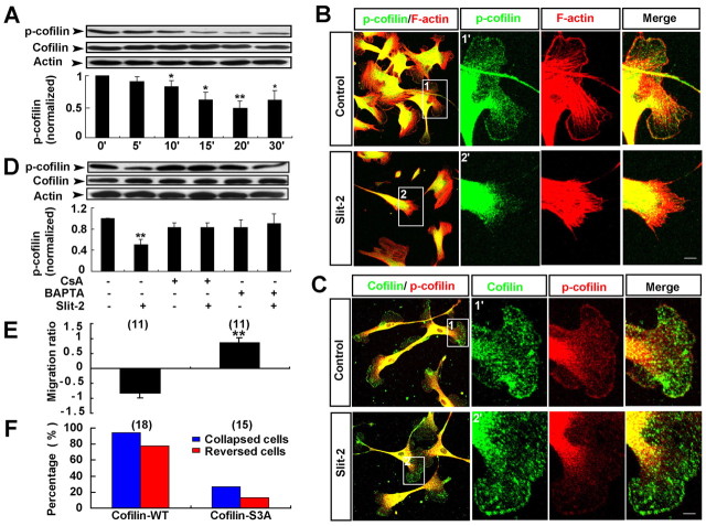
Activation of cofilin through Ca2+–calcineurin signaling is required for Slit-2-induced collapse and reversal of migration of OECs. (A) Western blot analysis for Slit-2-induced cofilin dephosphorylation and quantitative analysis of the ratio of p-cofilin to cofilin, normalized to the baseline level (0 minutes). (B) Immunocytochemical analysis of the distribution of p-cofilin (green) and F-actin (red) after Slit-2 treatment. Images of selected regions (1) and (2) are shown as (1′) and (2′) at higher magnification. (C) Immunocytochemical analysis of the distribution of cofilin (green) and p-cofilin (red) after Slit-2 treatment. (D) BAPTA-AM or CsA blocked the cofilin dephosphorylation triggered by Slit-2. (E,F) Average migration ratios (E) and the percentages of collapsed or reversed cells (F) in response to Slit-2 in total observed OECs transfected with GFP–cofilin-WT or GFP–cofilin-S3A. Data are mean + s.e.m.; *P<0.05, **P<0.01, Student's t-test. Scale bars: 20 μm.

