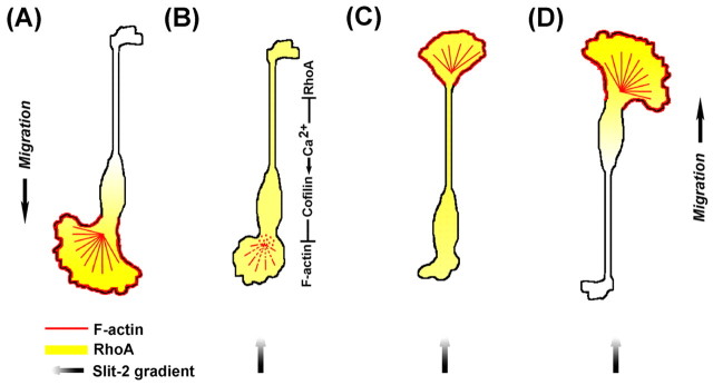
Fig. 9.

Model of Slit-2-induced collapse and reversal of migration of OECs. (A) Spontaneously migrating OEC exhibits a front–rear gradient in the distribution of RhoA. (B–D) Frontal exposure of a Slit-2 gradient triggers an elevation of [Ca2+]i at the leading front and soma, leading to the activation of cofilin, whose activity is responsible for the disruption of F-actin and the collapse of leading front. Reversal of the polarity of OECs requires both the gradient of RhoA inhibition across the cell and the collapse of the leading front. Colors indicate F-actin (red), RhoA (yellow) and Slit-2 gradient (black).
