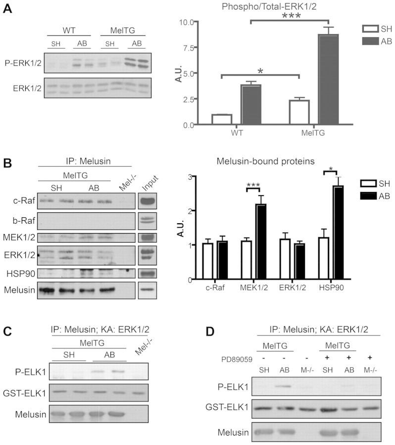
Fig. 1.
Melusin-bound MAPKs are activated by aortic banding. (A) Western blot analysis of phosphorylated and total ERK1/2 in hearts from wild-type (WT) and melusin-overexpressing (MelTG) mice in basal condition (SH; sham operated) and after 10 minutes of aortic banding (AB). The graph shows densitometric quantification of western blot bands (n=6 mice/group). (B) Immunoprecipitation of melusin from MelTG hearts after AB for 10 minutes or sham operation. Melusin-null hearts were used as negative controls. Co-immunoprecipitated proteins were visualized by western blot analysis. Input: heart total protein extract loaded on the same western blot as reference for molecular masses. Quantification of the co-precipitated proteins is shown in the graph (n=10/group). (C) ERK1/2 kinase assays were performed using GST–ELK1 as a substrate on melusin immunocomplexes obtained from MelTG hearts after AB for 10 minutes or sham operation. ERK1/2 kinase activity was revealed by western blot analysis with anti-phosphorylated ELK1 (Ser338; n=6/group). (D) ERK1/2 kinase assay performed on melusin immunocomplex in absence or presence of MEK1/2 inhibitor PD89059 (1 μM; n=3/group). *P<0.05; ***P<0.001; IP, immunoprecipitation; KA, kinase assay.

