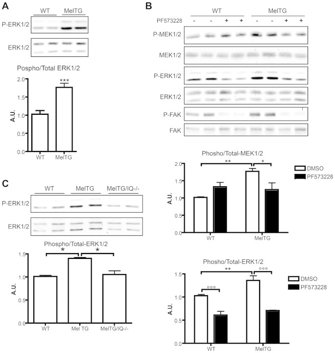
Fig. 4.
Melusin enhances ERK1/2 phosphorylation through FAK and IQGAP1. (A) Western blot analysis of phosphorylated and total ERK1/2 in neonatal cardiomyocytes obtained from wild-type and MelTG mice. The graph shows the densitometric quantification of western blot bands (n=5/group). (B) Phosphorylation and total protein levels of MEK1/2, ERK1/2 and FAK measured by western blotting on wild-type and MelTG cardiomyocytes untreated or treated with 3 μM PF573228. Anti-phosphorylated FAK (Tyr397), anti-phosphorylated MEK1/2 (Ser217/Ser221) and anti-phosphorylated ERK1/2 (Thr202/Tyr204) were used. Densitometric quantification of western blot bands is shown in the graph (n=4/group). (C) Western blot analysis of phosphorylated and total ERK1/2 on neonatal cardiomyocytes obtained from wild-type, MelTG and MelTG/Iqgap1-null (MelTg/IQ−/−) mice. The graph shows the densitometric quantification of western blot bands (n=4/group). *P<0.05; **P<0.01; ***P<0.001.

