Fig. 5.
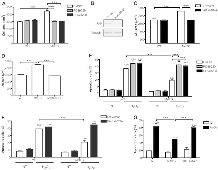
FAK and IQGAP1 are required for melusin-induced cardiomyocyte hypertrophy and survival. (A) The effect of the FAK inhibitor PF573228 and the MEK1/2 inhibitor PD89059 on cardiomyocyte surface areas, measured on at least 100 α-actinin-positive cells for each group (five cultures/group). (B) Representative western blots of FAK protein levels in cardiomyocytes infected with lentivirus encoding Fak shRNA or control viruses. Vinculin was used as a control for loading and RNA interference specificity. (C) Cardiomyocyte surface areas of cells treated as in B, measured on at least 100 α-actinin-positive, GFP-positive cells for each group (four cultures/group). (D) Wild-type, MelTG and MelTG/Iqgap1-null cardiomyocyte surface areas measured on at least 100 α-actinin-positive cells for each group (five cultures/group). (E) Percentage of apoptotic cardiomyocytes treated as in A, as indicated by TUNEL nuclear staining, measured on at least 100 α-actinin-positive cells for each group (five cultures/group). (F) Percentage of apoptotic cardiomyocytes treated as in B and C, as indicated by TUNEL nuclear staining and measured on at least 100 α-actinin-positive, GFP-positive cells for each group (4 cultures/group). (G) Percentage of apoptotic cardiomyocytes after treatment with H2O2, as indicated by TUNEL nuclear staining, measured on at least 100 α-actinin-positive cells for each group (five cultures/group). ***P<0.001; °°°P<0.001 versus untreated cells (NT).

