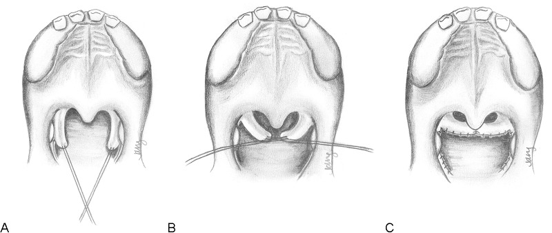
Fig. 5.

Technique for sphincter pharyngoplasty. (A) Musculomucosal flaps are elevated from the posterior tonsillar pillars on either side. Not shown: The uvula may be retracted for improved visualization. (B) Flaps are transposed into a horizontal direction to be inset into a transverse incision on the posterior pharyngeal wall. (C) The flaps are inset in an end-to-end fashion and the donor sites are sutured closed. The airway is smaller, but remains patent centrally. Note: For greater tightening of the sphincter, the flaps may be overlapped upon each other.
