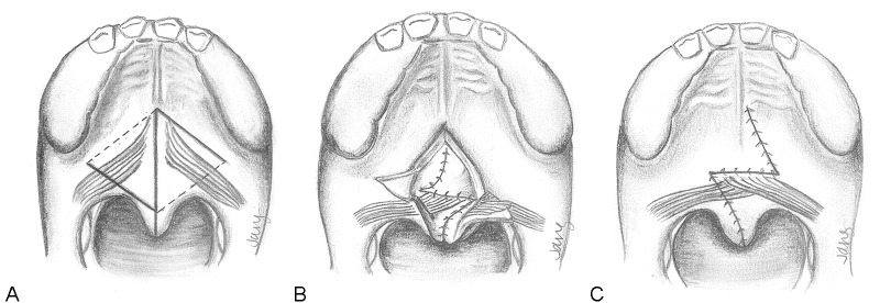
Fig. 6.

Technique for secondary Furlow palatoplasty. (A) The palate is divided at the midline. Oral mucosal incisions (dark lines) and nasal mucosal incisions (dotted lines) are shown. On the right side, an oral musculomucosal flap will be elevated, whereas the left oral flap will contain mucosa only. (B) The nasal Z-plasty has been transposed and this layer has been closed. Note that the left side contains the nasal myomucosal flap, which is now transversely oriented. The oral mucosa flaps remain elevated. (C) The oral mucosa is now closed. The palate has been lengthened by the Z-plasties and the levator musculature has been transposed and overlapped upon itself.
