
Fig. 5.
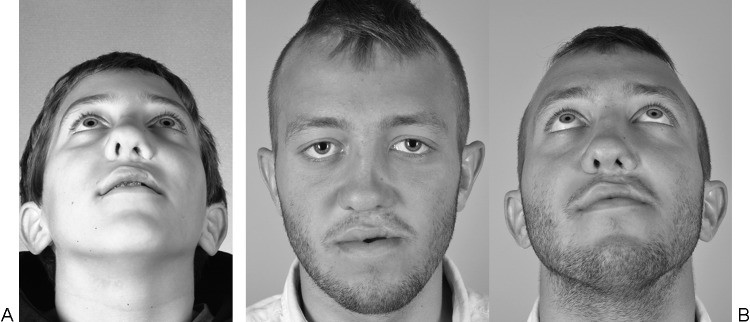
(A) Preoperative evaluation prior to the secondary rhinoplasty demonstrates a wide nasal root deviated away from the cleft with distorted cleft-side lower lateral cartilages. (B) Secondary rhinoplasty postoperative photos demonstrating corrected nasal tip projection and ala repositioning.