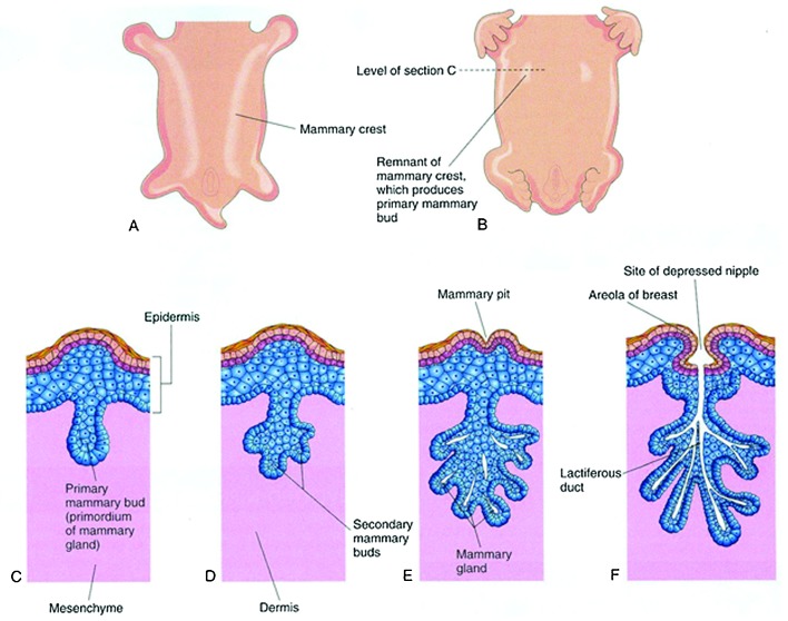
Fig. 1.
Development of the mammary gland. (A) Ventral view of an embryo at 28-days gestation showing mammary crests. (B) Similar view at 6-week gestation showing the remains of the mammary crests. (C) Transverse section of a mammary crest at the site of the developing mammary gland. (D–F) Similar sections showing successive stages of breast development between the 12th week of gestation and birth. (Reprinted with permission from Moore KL, Persaud TVN, Torchia MG, The Developing Human: Clinically Oriented Embryology. 9th ed. 2013 Copyright Elsevier).

