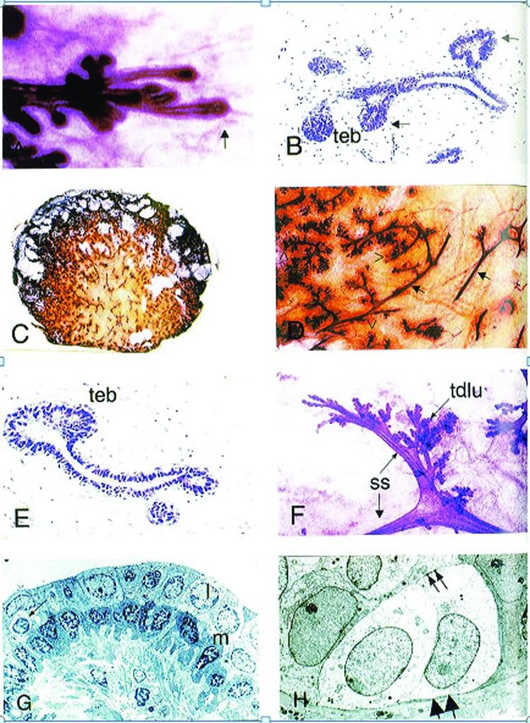
Fig. 3.
Pubertal breast development. (A) Carmine-stained whole-mount preparation of the advancing edge (arrow) of the parenchyma from a 13-year-old girl. (B) Hematoxylin- and eosin-stained developing breast of 13-year-old girl showing solid end bud-like structures (denoted teb) and lateral buds (arrows). (C) Coronal section of breast of 15-year-old girl. (D) Higher power view of panel C, arrows indicate ducts and unfilled arrowheads indicate duct termini. (E) Histology section of the peripheral region of parenchyma seen in (C). teb denotes terminal end bud. (F) Carmine-stained whole mount preparation of breast from 18-year-old nulliparous woman. A segmental duct divides into two subsegmental ducts (ss), which then lead to the terminal duct lobular units (tdlu). (G) Electron micrograph of a normal adult subsegmental duct. The bilayered histology with paler luminal cells (l), darker basal (myoepithelial) cells (m) is evident. An intraepithelial lymphocyte (arrow) is also seen. (H) Electron micrograph of a terminal duct lobular unit showing two basal clear cells. These have microfilaments in the basal part of the cell (large arrows) and desmosome attachments with the luminal cells (small arrows). (Reprinted with permission from Howard BA, Gusterson BA. Human breast development. J Mammary Gland Biol Neoplasia 2000;5(2):119–137. 2000 Copyright Springer).

