
Figure 1.
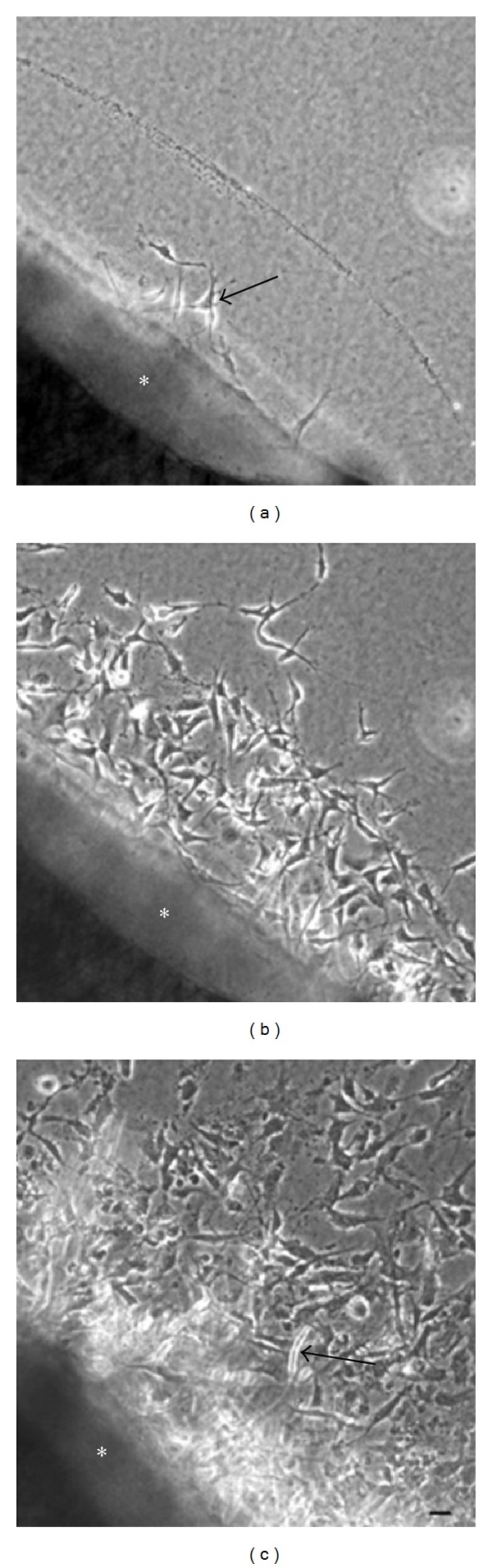
Phase-contrast microscopy. (a) After 2 days of cultivation, phase-contrast microscopy revealed fibroblastic cells (arrow) outgrown from a mouse aortic explant (∗) into a three-dimensional collagen gel. (b) After 5 days of cultivation, phase-contrast microscopy showed numerous fibroblastic cells outgrown from an aortic explant (∗). (c) After 7 days of cultivation, phase-contrast microscopy showed a tubular structure protruding (arrow) from an aortic explant (∗) into a three-dimensional collagen gel. (a), (b), and (c): Scale bar = 20 μm.