
Figure 2.
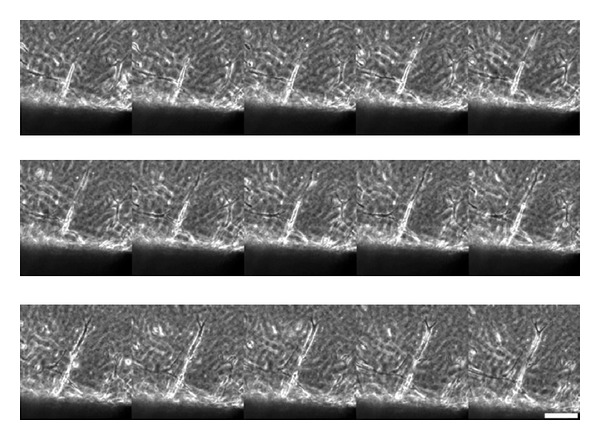
Time-lapse imaging of additional sprouts emerging and extending in a sequential manner from the leading edges of a newly formed capillary tube from the mouse aortic explant. Selected sequence from a time-lapse movie focusing on a single sprout. Note the protrusion of lamellipodia and continued migration of the leading cell. Scale bar = 50 μm.