
Figure 7.
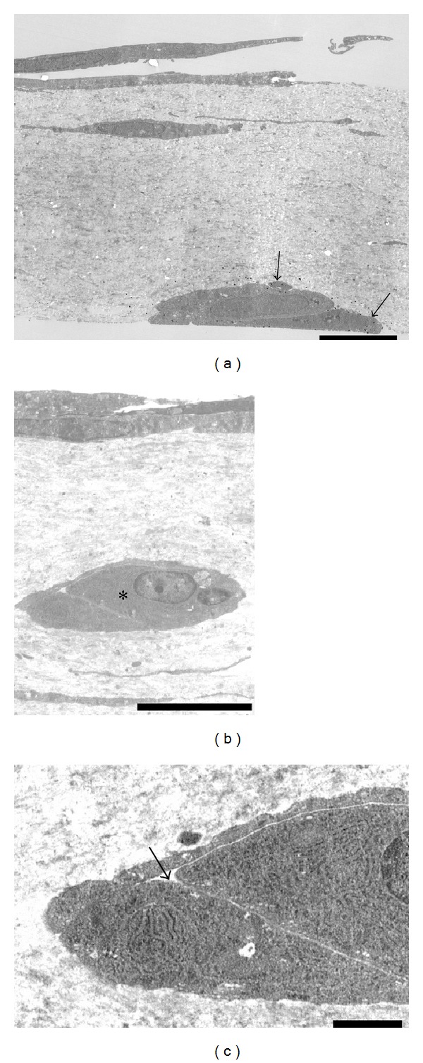
Immunoelectron microscopy of CD133. (a) At the bottom side of the collagen gels, CD133-positive cells made contact with each other (arrows). Cell organelles were sparse in the cells. Scale bar = 5 μm. (b) CD133-negative cells made contact with each other in the middle layer of the collagen gels (∗). Scale bar = 5 μm. (c) Enlargement of (b) (∗). These cells formed intercellular vacuolar structures (arrow). Cell organelles, such as the rough endoplasmic reticulum, were rich in these cells. Scale bar = 1 μm.