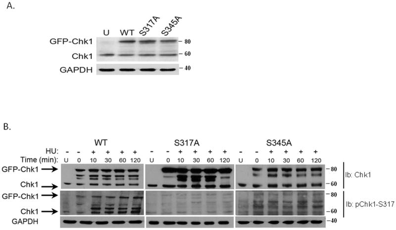
Fig. 4. Analysis of Serine phosphorylation sites in Chk1 in response to HU.

A. Recombinant wild type pEGFP-Chk1 and the phosphorylation mutants: pEGFP-Chk1-S317A and pEGFP-Chk1-S345A plasmids were transiently transfected into MCF7 cells and expression of endogenous and recombinant proteins was detected by western blot analysis.
B. MCF7 cells were transiently transfected with wild type or phosphorylation mutants of Chk1 and 24 hours after transfection, cells were treated with 2 mM HU for the indicated time points. The expression and phosphorylation levels of Chk1 were examined by western blot analysis with phospho-Chk1-specific antibodies and anti-Chk1 antibody.
