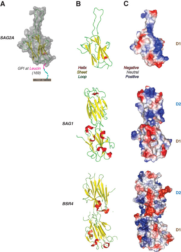
Figure 2.
Anchorage site, carbon structure and charge distribution of SAG2A protein. (A) SAG2A anchorage in Toxoplasma gondii’s surface surface by glycosyl-phosphatidylinositol (GPI) was predicted to be in a leucin at position 169 of its amino acid sequence, located at the C-terminal end of SAG2A. (B) The modeled carbon structure of SAG2A evidences a disordered amino acid sequence, absent in the SAG1 and BSR4 proteins. (C) The C-terminal end of SAG2A presents a relevant hydrophobic portion, with distinct polar amino acids at positions 134 and 137.

