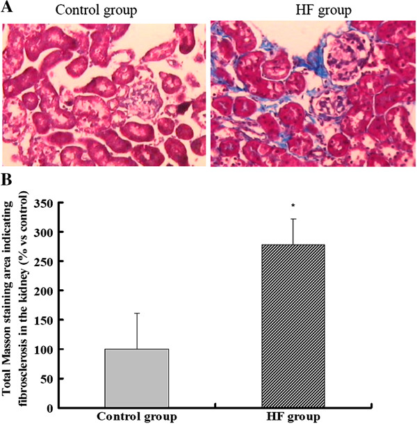
Figure 1.

Morphological changes in the kidneys of ApoE KO mice. (A) Masson staining showed significant deposition of collagen in the tubular interstitium of the HF group when compared with the control group (×200). (B) Quantitative analysis of fibrotic tissue stained by Masson staining. Positive stainging was quantified by image analysis using Image J software by a point-counting technique under a 176-point grid. The histogram represents the mean ± SD of the percentage of the field area from eight experiments. * P < 0.05 vs. control.
