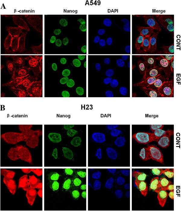
Figure 4.
Expression of β-catenin and Nanog was concomitantly regulated by EGFR signaling. A549 (A) and H23 (B) cells were treated without or with EGF followed by immunofluorescent staining. In the absence of EGF, β-catenin was located predominantly at the plasma membrane, with faint staining distributed in the cytoplasm. Faint Nanog staining was detected in the nucleus. When cells were treated with EGF, β-catenin staining translocated to the nucleus in both cell lines and nuclear Nanog staining was moderately increased in A549 cells and abundantly accumulated in H23 cells.

