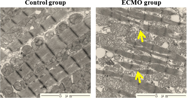
Figure 3.

Ultrastructure of cardiomyocyte under TEM (magnification*8000). Ultrastructure of cardiomyocyte was normal in the control group, and mildly disorderd of myofilament and dissolved of focal myofilament (pointed by arrow) were observed in the ECMO group.
