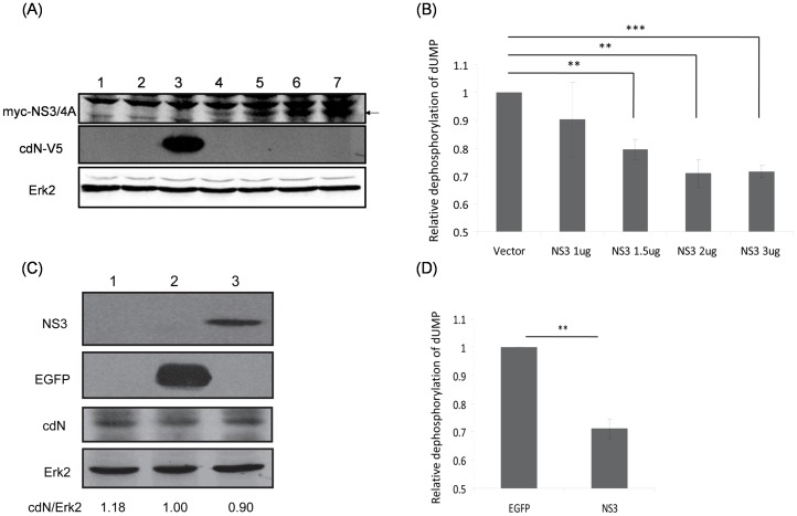
Figure 5. HCV NS3 protein partially represses cellular cdN activity.
(A) HuH7 cells were mock-transfected (lane 1) or transfected with empty vector (3 ug, lane 2), the cdN plasmid (3 ug, lane 3) or different amount of myc-NS3/4A plasmids (1 ug, lane 4; 1.5 ug, lane 5; 2 ug, lane 6; 3 ug, lane 7) together with empty vectors to a total of 3 ug DNA in each experiment. At 48 hrs after transfection, proteins derived from these cells were analyzed using antibodies against myc tag to detect the expression of exogenous NS3/4A protein (upper panel), against V5 tag to detect the exogenous cdN expression (middle panel) or against Erk-2 as a loading control (bottom panel). (B) The 5′(3′)-deoxyribonucleotidase activity was measured using cell lysates derived from (A). (C) HuH7 cells were mock-transduced (lane 1) or transduced with lentiviral vectors expressing EGFP (lane 2) or HCV NS3/4A protein (lane 3). After puromycin selection, proteins derived from these cells were analyzed using antibodies against NS3 (upper panel), against EGFP, against cdN protein or against Erk-2 as a loading control (bottom panel). (D) The 5′(3′)-deoxyribonucleotidase activity was analyzed using cell lysates derived from (C).

