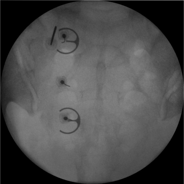
Figure 2.

Anteroposterior fluoroscopic view of the sacrum showing three separate 25-gauge spinal needles placed within the left S1, S2, and S3 foramina. An epsilon ruler is used as a guide such that the needle tip is positioned about 10 mm lateral to the posterior sacral foramina apertures. The radiofrequency electrode is positioned at the 11 o’clock position of the S1 foramen.
