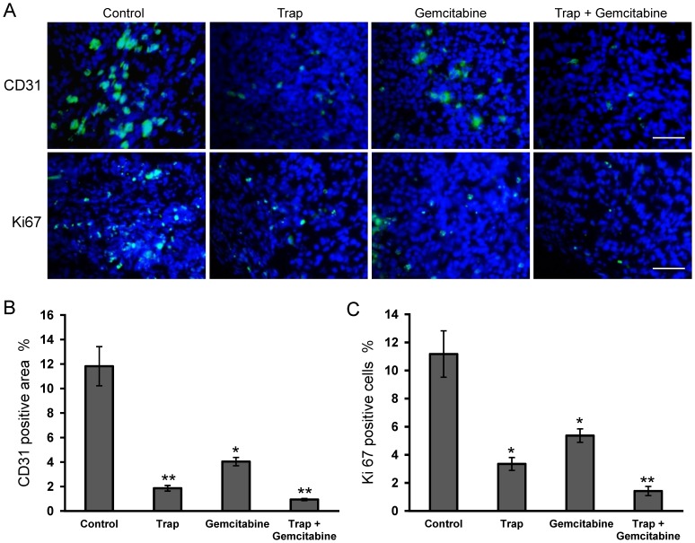
Figure 3. Inhibition of angiogenesis and cell proliferation within tumors after the combination therapy.
(A) Representative images of CD31-positive microvessel area and Ki67-positive cells in the viable LLC tumor tissues on day 15 after treatment initiation were estimated by immunohistochemical staining; (B) and (C) Microvessel density and percentage of Ki67-positive cells were determined by counting the number of the positive staining per high-power field in the section, as described in “Materials and Methods”. *P<0.05, **P<0.01 vs the control group. Scale bar, 50 µm.

