
Figure 1. SHP protein is localized on the mitochondrial outer membrane.
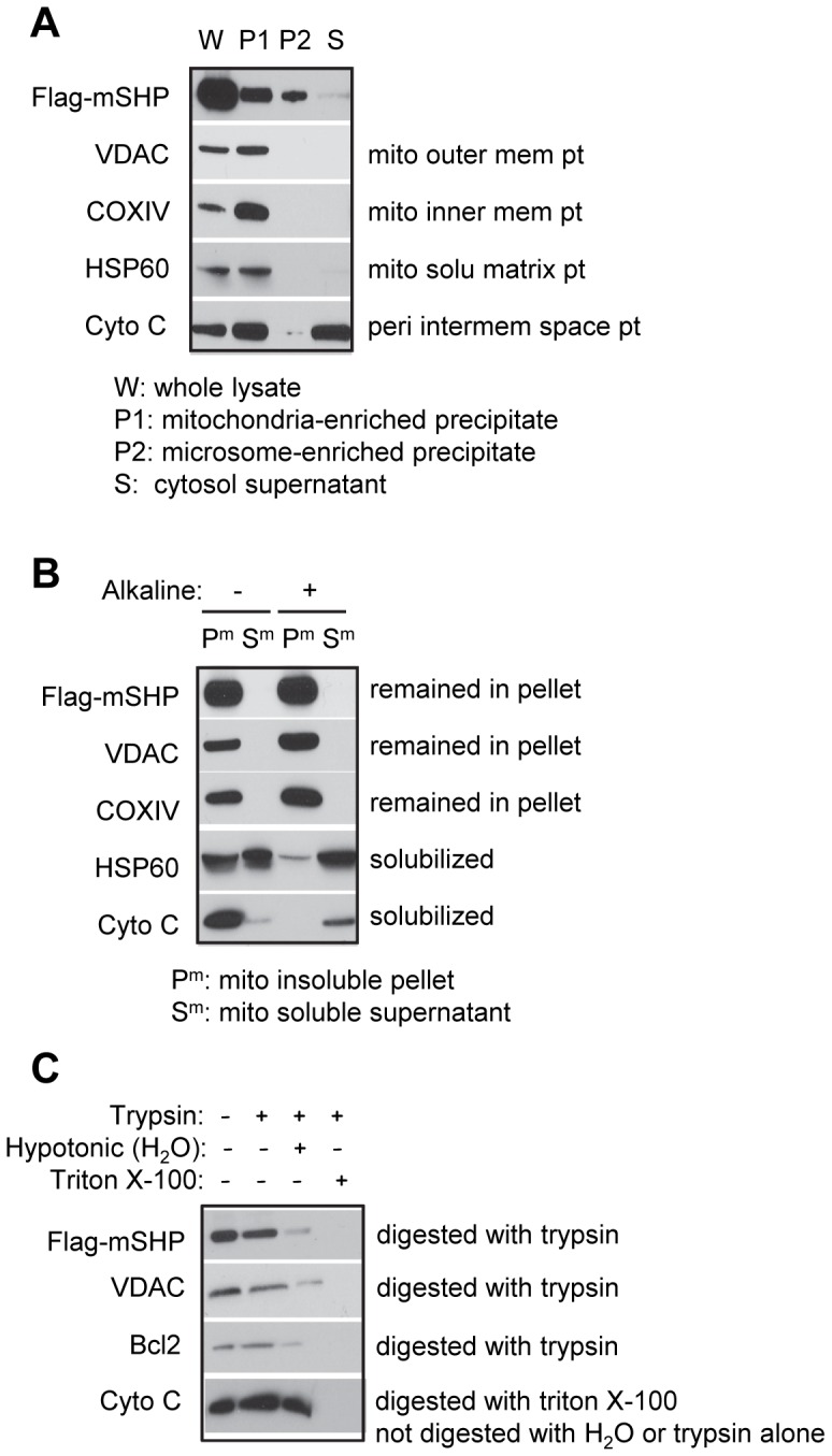
(A) Western blots to detect Flag-mSHP, VADC, COXIV, HSP60, and Cyto C proteins using specific antibodies for each protein, with the exception of SHP, which was detected with an anti-Flag antibody. Huh7 cells were transfected with Flag-mSHP (20 µg, 15 cm plate), and five plates were used to harvest protein. Differential centrifugation was used to isolate the mitochondria-enriched precipitate fraction (P1), microsome-enriched precipitate fraction (P2), and cytosolic supernatant fraction (S); the whole cell lysate (W) was used for comparison. (B) Alkaline digestion of mitochondrial membrane fraction (Pm) and soluble fraction (Sm) and Western blots to determine Flag-mSHP, VADC, COXIV, HSP60, and Cyto C proteins. (C) Trypsin protection assays and Western blots to determine Flag-mSHP, VADC, Bcl2, and Cyto C proteins in mitochondria. Abbreviations: mito, mitochondria; mem, membrane; solu, soluble; peri, peripheral; pt, protein.