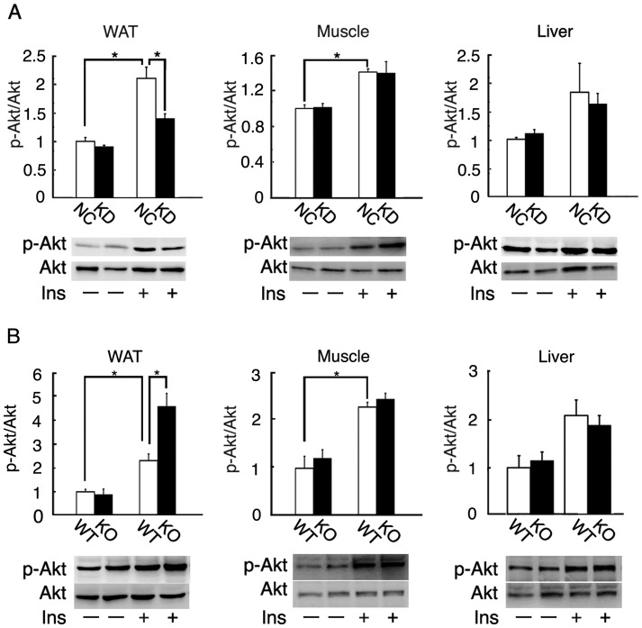
Figure 7. Insulin-stimulated Akt phosphorylation of wild-type and Fgf21 knockout mice.
(A) Basal or insulin-stimulated Akt phosphorylation in the subcutaneous white adipose tissue, gastrocnemius muscle, and liver of 3-month-old wild-type mice fed NC or KD for 6 days (n = 7 per group; *, P<0.05). (B) Basal or insulin-stimulated Akt phosphorylation in the subcutaneous white adipose tissue, gastrocnemius muscle, and liver of 3-month-old wild-type (WT) and Fgf21 knockout (KO) mice fed KD for 6 days (n = 7 per group; *, P<0.05). Akt and the phosphorylated form were detected by Western blotting with specific antibodies.

