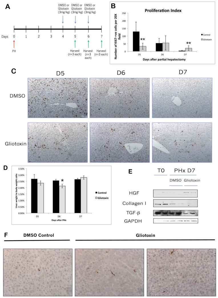
Figure 6. Treatment with gliotoxin during the later stages of liver regeneration results in increased proliferation and decreased matrix re-deposition.
(A) Schematic of the late-stage gliotoxin study, in which rats were subjected to PH followed by treatment with DMSO or 3mg/kg gliotoxin on days 4, 5, and 6 after PH. Livers were harvested 5, 6, or 7 days following PH. (B) Quantification of the Ki67 staining in (C). (C) Representative images of Ki67 IHC on livers harvested at D5, D6, and D7 after PH in gliotoxin and control groups (100X). (D) Graph of liver weight to body weight ratios after late DMSO/gliotoxin treatment. (E) WB analysis of whole cell lysates from control and gliotoxin-treated livers harvested at D7. (F) TUNEL staining on livers from the late-stage experiment (200X).

