Figure 4.
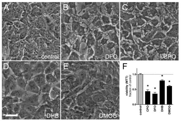
HIF-PHD inhibitors decrease the viability of DLD-1 cells. A-E, phase contrast images of A, untreated DLD-1 cells or cells treated with B, DFO 50 μM, C, CPO 1 μM, D, DHB 20 μM, or E, DMOG 250 μM for 72 h. CPO or DFO (B and C) caused the formation of large cytosolic vacuoles, while all compounds yielded larger cells. F, DLD-1 cultures were assayed for viability by MTT reduction following treatment with HIF-PHD inhibitors. Scale bar = 25 μm. *=P<0.01 vs. control, one-way ANOVA-Bonferroni’s post test, n=8.

