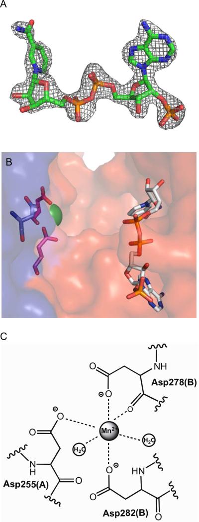
FIGURE 5.
“Ligands in the Mtb ICDH-1 Structure.” (A) Electron density map for NADPH at 2σ. (B) A close up of one of the two the active sites showing Mn2+ as a green sphere and NADPH as sticks. Coordinating residues of the Mn2+ from the A chain are shown in pink, and the residue from the B chain is shown in blue.
(C) Metal binding site.

