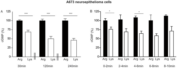
Figure 2. Preservation of residual nNOS activity also under prolonged arginine withdrawal in human A673 neuroepithelioma cells.
(A) NO measurements were performed indirectly by cGMP formation in RFL-6 reporter cells as described in Figure 1(A), except that the pre-incubation in LS containing either 1 mM arginine (dark columns) or 1 mM lysine (white columns) was for 30, 120, or 240 min as indicated and only one transfer each was performed after stimulation with calcium ionophore. After substraction of the basal cGMP content of the RFL-6 cells, values obtained from cells incubated in 1 mM lysine were calculated as % of the mean of the values obtained from the corresponding control cells incubated in 1 mM arginine. Columns represent mean ± S.E.M. (n = 9–12, unpaired student’s t-test). (B) Cells were preincubated in LS containing either 1 mM arginine (dark columns) or 1 mM lysine (white columns) for 120 min. NO measurements and data analyses were performed as in Figure 1 (A), except that the same A673 cells were assayed five times consecutively. After each transfer, fresh LS supplemented with the indicated amino acid and 10 µM calcium ionophore A23187 was added to the A673 cells, incubated for 2 min and transferred to new RFL-6 cells. The basal cGMP content of the RFL-6 cells was subtracted. The values obtained from cells incubated in 1 mM lysine were calculated as % of the mean of the values obtained from the corresponding control cells incubated in 1 mM arginine for 0–2 min. Columns represent mean ± S.E.M. (n = 6, one way ANOVA with Bonferroni post-hoc test).

