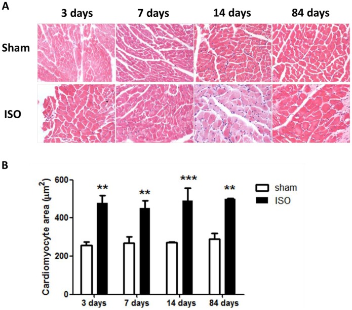
Figure 2. Isoproterenol induced cardiac hypertrophy as detected by HE staining.
(A) HE staining of sections from the left ventricular myocardium of mice subjected to vehicle or isoproterenol. (B) Quantification of cross-sectional areas of the cardiomyocytes shown in (A). **P<0.01, ***P<0.001, compared with sham at the same time points. ISO, isoproterenol.

