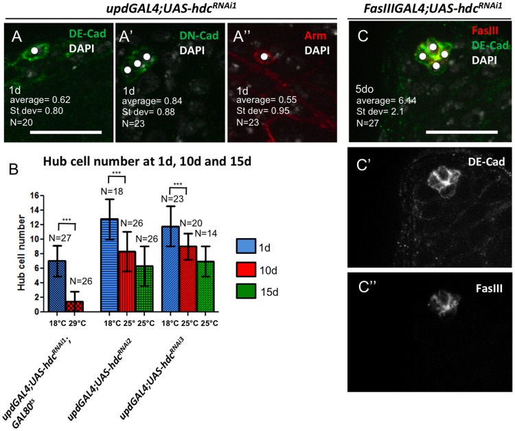
Figure 2. Hub cell loss is evident using multiple paradigms and is not due to developmental defects.
(A to A’”) Strong hub cell loss marked by staining for FasIII (see Fig. 1C and F) was confirmed with other hub cell markers [DE-Cadherin (DE-Cad), DN-Cadherin (DN-cad) and Armadillo (Arm)] Hub cells pointed by white dots. (B) Hub cell quantification in flies where hdcRNAi expression by updGal4 was suppressed at 18°C during development, and activated at 25°C (without Gal80ts; hdcRNAi2 and hdcRNAi3) or 29°C (with Gal80ts; hdcRNAi1) for 1, 10, and 15 days. Means and SD are shown; ***P<0.001 (Kruskal–Wallis one-way analysis of variance). (C) Loss of hub cells is observed using an alternative hub driver (FasIIIGal4). Testis from FasIIIGal4; UAS-hdcRNAi1 male at 5 days (compare to Fig. 1E); Scale bars 20 µm.

